How Can You Treat Bursitis Of The Foot?
Overview
Bursae (two or more bursa) are small, fluid-filled sacs that cushion the bones, tendons and muscles surrounding your joints. They contain a lubricating fluid that reduces friction, allowing tissues in the body to glide past each other smoothly. Imagine the bursa as a protective layer that helps keep a tendon or muscle from fraying or getting aggravated as it eases over a bone or around a corner. Bursitis is a condition that occurs when a bursa becomes inflamed: irritated, red and filled with more fluid than normal.
Causes
There are several factors which can lead to a person developing retrocalcaneal bursitis. In athletes, especially runners, overtraining, sudden excessive increase in running mileage may lead to retrocalcaneal bursitis. Tight or ill-fitting shoes can be another causative factor as they can produce excessive pressure at the back of the heel due to restrictive heel counter. A person with an excessively prominent posterosuperior aspect of the heel bone (Haglund deformity) may also have a higher predisposition to retrocalcaneal bursitis. In such individuals, pain would be reproduced when the ankle goes into dorsiflexion.
Symptoms
Retrocalcaneal bursitis is very similar to Achilles bursitis as the bursae are very close in proximity and symptoms are almost identical however retrocalcaneal bursitis is a lot more common. The symptoms of bursitis vary depending on whether the bursitis is the result of injury or an underlying health condition or from infection. From normal overuse and injury the pain is normally a constant dull ache or burning pain at the back of the heel that is aggravated by any touch, pressure like tight shoes or movement of the joint. There will normally be notable swelling around the back of the heel. In other cases where the bursa lies deep under the skin in the hip or shoulder, swelling might not be visible. Movement of the ankle and foot will be stiff, especially in the mornings and after any activity involving the elbow. All of these symptoms are experienced with septic bursitis with the addition of a high temperature of 38?C or over and feverish chills. The skin around the affected joint will also appear to be red and will feel incredibly warm to the touch. In cases of septic bursitis it is important that you seek medical attention. With injury induced bursitis if symptoms are still persisting after 2 weeks then report to your GP.
Diagnosis
A thorough subjective and objective examination from a physiotherapist may be all that is necessary to diagnose a retrocalcaneal bursitis. Diagnosis may be confirmed with an ultrasound investigation, MRI or CT scan.
Non Surgical Treatment
In addition to R.I.C.E., there are a number of other treatments to reduce swelling and any associated pain or discomfort due to heel bursitis. Orthotics or change of footwear. Wearing an orthotic device such as a heel insert can encourage better mechanics in the foot and reduce irritation of the retrocalcaneal bursa. Some people do not need special orthotics but simply need to stop wearing shoes with rigid heel and ankle construction and instead wear more supportive, comfortable shoes. Shoes with an "Achilles notch," a groove in the collar at the back of the shoe to protect the Achilles tendon, can be particularly helpful. (Almost all running shoes are designed with an Achilles notch.) Stretching and physical therapy. Stretching the Achilles tendon often helps alleviate pain. Once the pain is resolved it is important for the patient to continue a regular stretching program. Regular stretching reduces the chance of recurrence.
Surgical Treatment
Surgery to remove the damaged bursa may be performed in extreme cases. If the bursitis is caused by an infection, then additional treatment is needed. Septic bursitis is caused by the presence of a pus-forming organism, usually staphylococcus aureus. This is confirmed by examining a sample of the fluid in the bursa and requires treatment with antibiotics taken by mouth, injected into a muscle or into a vein (intravenously). The bursa will also need to be drained by needle two or three times over the first week of treatment. When a patient has such a serious infection, there may be underlying causes. There could be undiscovered diabetes, or an inefficient immune system caused by human immunodeficiency virus infection (HIV).
Flexible Hammer Toe Therapy
 Overview
What Is A Hammer toe? A hammer toe, or claw toe, describes a condition where the toe(s) become buckled, contracted or crooked. The toe could even cross over an adjacent toe, which is called a cross over toe. Any of the toes may be affected, but the 2nd and 5th toe are most commonly involved.
Causes
While there are a number of causes, there aren't many specific risk factors for hammertoes, women tend to get these problems more than men, but they occur without rhyme or reason. Diabetics, however, are more likely to get a hammertoe if they have underlying nerve damage in the toes and feet.
.jpg) Symptoms
Symptoms
Hammer, claw, and mallet toes can cause discomfort and pain and may make it hard to walk. Shoes may rub on your toes, causing pain, blisters, calluses or corns, or sores. Sores can become infected and lead to cellulitis or osteomyelitis, especially if you have diabetes or peripheral arterial disease. If you have one of these health problems and sores develop, contact your doctor.
Diagnosis
Hammertoes are progressive, they don?t go away by themselves and usually they will get worse over time. However, not all cases are alike, some hammertoes progress more rapidly than others. Once your foot and ankle surgeon has evaluated your hammertoes, a treatment plan can be developed that is suited to your needs.
Non Surgical Treatment
Treating hammertoe involves straightening the toe, making tendons in the toes flexible again, and preventing the problem from returning. Some simple treatments include Soaking your feet every day in warm water, then stretching your toes and ankles by pointing your toes. Using over-the-counter pads, cushions or straps to decrease discomfort. Splinting the toe to keep it straight and to stretch the tendons of the foot. Exercising the toes to relax the foot tendons (a session with a physical therapist may help you get started with foot exercises). One simple exercise is to place a small towel on the floor and then pick it up using only your toes. You also can grasp at carpet with your toes or curl your toes up and down repeatedly. Wearing shoes that fit properly and give toes plenty of room to stretch out.
Surgical Treatment
Surgery is the approach that is often necessary to correct hammertoe that fails to respond to nonsurgical management. Surgery is appropriate when the muscles and tendons involved in a hammertoe problem have become so tight that the joints are rigid, misaligned and unmovable. There are a number of surgical techniques for dealing with the complex range of joint, bone, muscle, tendon and ligament abnormalities that define each hammertoe's make-up. To correct a hammertoe deformity, the surgeon's goal is to restore the normal alignment of the Hammer toes toe joint, relieving the pressure that led to the hammertoe's development (this should also relieve the pain, as well). To do this, he or she may remove part of the boney structure that creates a prominence at the top of the joint. Tighten or loosen the muscles, tendons and ligaments around the toe joints. Realign the toe bones by cutting one or more and shifting their position, realigning muscles, tendons and ligaments accordingly. Use screws, wires or plates to hold the joint surfaces together until they heal. Reconstruct a badly damaged joint or replace it with an artificial implant.
What Causes Bunions?
Overview
 A bunion, or Hallux Valgus, is a foot deformity characterised by deviation of the bones around the big toe joint of the foot. As a result, there is a large exostosis or bony lump on the inside of the foot and the toe is pointed across towards the smaller toes. It is a common problem, more so in women and has been attributed to tight fitting footwear. This article will cover the basics of bunions, what it is due to, and what treatment is available.
Causes
Women are particularly at risk for bunions, as high heels may help aggravate the condition. Bunions may also be caused when bursal sacs, sacs that absorb friction and lubricate the many tendons, bones muscles of the body, in the foot become inflamed or swollen. Bunions may also be prompted by the metatarsal bone, the bone that runs from the heel to the toe of the foot, with one metatarsal bone leading to each toe, of the big toe enlarging or deviating slightly to the outside of the foot.
Symptoms
While bunions may be considered cosmetically undesirable, they are not necessarily painful. In cases where the individual has minor discomfort that can be eased by wearing wider shoes made of soft leather and/or with the aid of spacers-padding placed between the toes to correct alignment-further treatment may not be necessary. (Anti-inflammatory agents can be used to alleviate temporary discomfort at the site of the bursa.) For those who continue to experience pain on a daily basis and who cannot wear most types of shoe comfortably, surgical treatment may be the best choice.
Diagnosis
A doctor can very often diagnose a bunion by looking at it. A foot x-ray can show an abnormal angle between the big toe and the foot. In some cases, arthritis may also be seen.
Non Surgical Treatment
The initial treatment of a bunion should be non-operative. Symptoms can often be greatly improved with simple non-operative interventions. Non-operative treatment may include properly fitted shoes, Properly fitting comfort shoes with a wide non-constrictive toe box, especially one that is made out of a soft material such as leather, can be quite helpful in reducing the irritation over the prominent bunion. In some instances, it is helpful to have a shoemaker stretch the inside aspect of the shoe. Jamming a foot with a bunion into a constrictive shoe will likely lead to the development of uncomfortable symptoms. Bunion pads, Medial bunion pads may also be helpful in decreasing the symptoms associated with the bunion. These pads can be obtained at many drugstores. Essentially, they serve to lessen the irritation over the medial prominence and, thereby, decrease the associated inflammation This should be combined with comfortable non-constrictive shoes. A toe spacer placed between the great toe and the second toe can help to reduce the bunion deformity and, thereby, decrease the stretch on the medial tissue and the irritation associated with the bunion. Toe spacers can be obtained at most drug stores or online. Soft shoe inserts. Over-the-counter accommodative orthotics may also help bunion symptoms. This product is particularly helpful if bunion symptoms include pain that is under the ball of the foot. Orthotics with a slight medial longitudinal arch may be helpful for patients that have associated flatfoot deformity. These can be purchased at many sports stores, outdoors stores, or pharmacies. Bunion splints have often been used to treat the symptoms associated with hallux valgus. These splints are typically worn at night in an effort to reduce the bunion deformity. There is no evidence to suggest that these splints decrease the rate at which bunion deformities occur. There is also no evidence that clearly supports their effectiveness. However, some patients report good relief with the use of these splints. 
Surgical Treatment
Surgery takes place either under local or general anaesthetic and takes about one hour. After surgery you will have either a plaster cast or special dressing on the foot and you will be given a special walking shoe and crutches to use the first few days/weeks. Recovery usually takes approximately 6-8 weeks but swelling often lasts longer and it may take a few months before you are able to wear normal shoes again. Full recovery can take up to a year. Bunion surgery is successful in approximately 85% of cases, but it is vital not to go back to wearing ill-fitting shoes else the problem is likely to return.
Prevention
To help prevent bunions, select your style and size of shoes wisely. Choose shoes with a wide toe area and a half-inch of space between the tip of your longest toe and the end of the shoe. Shoes also should conform to the shape of your feet without causing too much pressure.
Over-Pronation
Overview
Too much of a good thing can be bad, and when it comes to pronation of the foot this is often the case. When the foot rolls too much during contact with the ground, the body weight is not cushioned effectively. The degree to which pronation occurs may be difficult to spot without someone observing how you run. It is easier to tell if a runner is pronating excessively when they are observed from behind rather than from above. When overpronation is only mild to moderate it may not be easy to spot without having a professional gait analysis performed.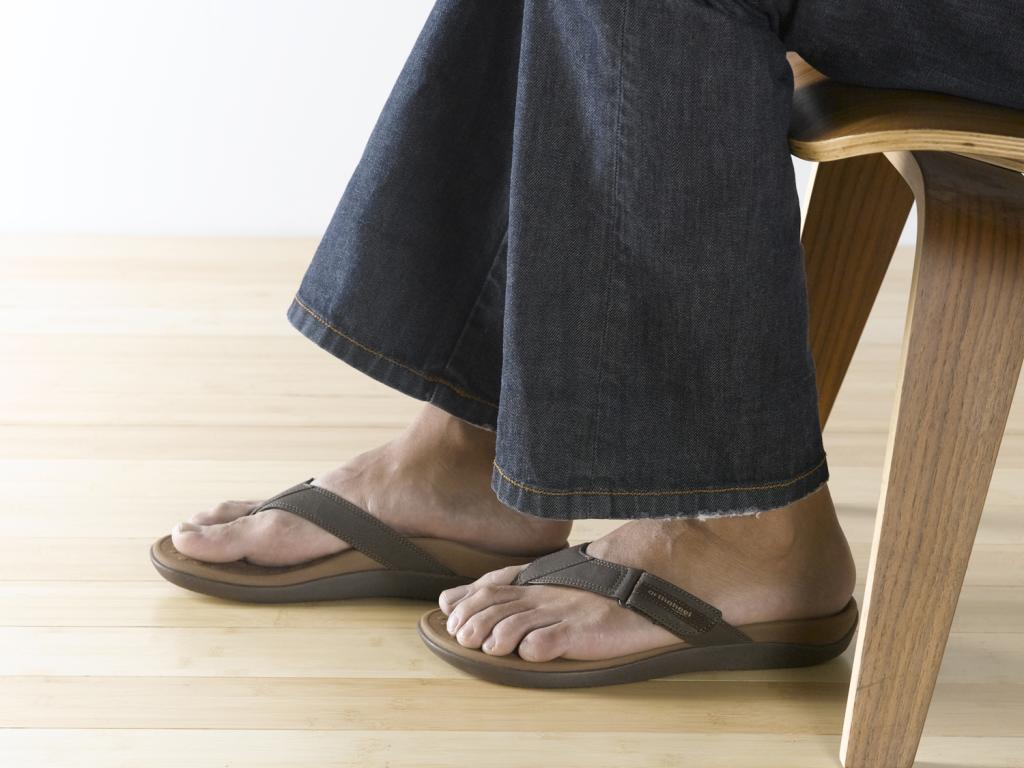
Causes
Over-pronation may happen because the tissue that attaches to your foot bones is loose. You may be born with this problem or it may result from injuries or overuse, like from too much running.
Symptoms
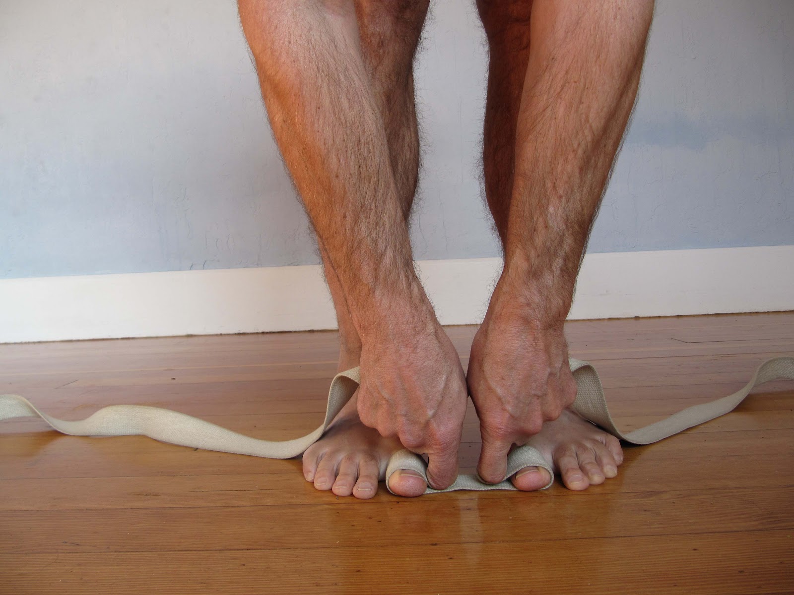
Over-pronation is a condition where the arch flattens out which makes the feet roll inward while walking. This condition is also known as flat feet. It imposes extreme additional stresses on the plantar fascia, a fibrous band of tissue which connects the heel to the forefoot. Over-pronation makes walking a painful experience because of the additional strain on the calves, heel and/or back. Treatment for over-pronation involves the use of specially-made orthotics which offers arch support and medial rear foot posting as corrective measures.
Diagnosis
If you have flat feet or low arches, chances are you overpronate. Although not always the case, the lower your arches the greater the overpronate. Stand on a hard surface (in front of a mirror if you need to) and look at your feet, flat feet or low arches are easy to spot. If your feet look flatter than a pancake, have a look at your ankles and see if they seem collapsed or straight. If they are, you're overpronating.
Non Surgical Treatment
Wear shoes with straight or semicurved lasts. Motion-control or stability shoes with firm, multidensity midsoles and external control features that limit pronation are best. Over-the-counter orthotics or arch supports can help, too. You know you are making improvements when the wear pattern on your shoes becomes more normal. Overpronation causes extra stress and tightness to the muscles, so do a little extra stretching.
Surgical Treatment
Depending on the severity of your condition, your surgeon may recommend one or more treatment options. Ultimately, however, it's YOUR decision as to which makes the most sense to you. There are many resources available online and elsewhere for you to research the various options and make an informed decision.
Too much of a good thing can be bad, and when it comes to pronation of the foot this is often the case. When the foot rolls too much during contact with the ground, the body weight is not cushioned effectively. The degree to which pronation occurs may be difficult to spot without someone observing how you run. It is easier to tell if a runner is pronating excessively when they are observed from behind rather than from above. When overpronation is only mild to moderate it may not be easy to spot without having a professional gait analysis performed.
Causes
Over-pronation may happen because the tissue that attaches to your foot bones is loose. You may be born with this problem or it may result from injuries or overuse, like from too much running.
Symptoms
Over-pronation is a condition where the arch flattens out which makes the feet roll inward while walking. This condition is also known as flat feet. It imposes extreme additional stresses on the plantar fascia, a fibrous band of tissue which connects the heel to the forefoot. Over-pronation makes walking a painful experience because of the additional strain on the calves, heel and/or back. Treatment for over-pronation involves the use of specially-made orthotics which offers arch support and medial rear foot posting as corrective measures.
Diagnosis
If you have flat feet or low arches, chances are you overpronate. Although not always the case, the lower your arches the greater the overpronate. Stand on a hard surface (in front of a mirror if you need to) and look at your feet, flat feet or low arches are easy to spot. If your feet look flatter than a pancake, have a look at your ankles and see if they seem collapsed or straight. If they are, you're overpronating.
Non Surgical Treatment
Wear shoes with straight or semicurved lasts. Motion-control or stability shoes with firm, multidensity midsoles and external control features that limit pronation are best. Over-the-counter orthotics or arch supports can help, too. You know you are making improvements when the wear pattern on your shoes becomes more normal. Overpronation causes extra stress and tightness to the muscles, so do a little extra stretching.
Surgical Treatment
Depending on the severity of your condition, your surgeon may recommend one or more treatment options. Ultimately, however, it's YOUR decision as to which makes the most sense to you. There are many resources available online and elsewhere for you to research the various options and make an informed decision.
What Are The Primary Causes Of Severs Disease?
Overview
Sever's disease (calcaneal apophysitis) is the term used to describe irritation (inflammation) of the calcaneal apophysis. This condition often occurs before or during the growth spurt in boys and girls, or shortly after they begin a new activity. Sever's disease is common is running and jumping sports.
Causes
Sever's disease usually develops as a result of overuse and is common in active children between the ages of 8 to 12. Activities that involve running or jumping can cause undue stress on the calcaneal apophysis. This in turn leads to the development of microscopic damage to the calcaneal apophysis resulting in inflammation and pain. Poor flexibility of the calf muscles and of the Achilles tendon, overpronation (feet rolled in) and inappropriate footwear are some of the other factors that can cause Sever's disease.
Symptoms
The most obvious sign of Sever's disease is pain or tenderness in one or both heels, usually at the back. The pain also might extend to the sides and bottom of the heel, ending near the arch of the foot. A child also may have these related problems, swelling and redness in the heel, difficulty walking, discomfort or stiffness in the feet upon awaking, discomfort when the heel is squeezed on both sides, an unusual walk, such as walking with a limp or on tiptoes to avoid putting pressure on the heel. Symptoms are usually worse during or after activity and get better with rest.
Diagnosis
A doctor or other health professional such as a physiotherapist can diagnose Sever?s disease by asking the young person to describe their symptoms and by conducting a physical examination. In some instances, an x-ray may be necessary to rule out other causes of heel pain, such as heel fractures. Sever?s disease does not show on an x-ray because the damage is in the cartilage.
Non Surgical Treatment
Treatment may consist of one or more of the following. Elevating the heel. Stretching hamstring and calf muscles 2-3 times daily. Using R.I.C.E. (Rest, Ice, Compression, Elevation). Foot orthotics. Medication. Physical therapy. Icing daily (morning). Heating therapy. Open back shoe are best and avoid high heel shoe. The Strickland Protocol has shown a positive response in patients with a mean return to sport in less than 3 weeks. Further research into the anatomical and biomechanical responses of this protocol are currently being undertaken.
Recovery
With proper care, your child should feel better within 2 weeks to 2 months. Your child can start playing sports again only when the heel pain is gone. Your doctor will let you know when physical activity is safe.
Posterior Tibial Tendon Dysfunction
Overview
Adult Acquired Flatfoot occurs when the arch of your foot collapses after your skeleton has stopped growing, usually resulting in the foot ?falling? inward with the toes pointing out. This allows your entire sole to touch the ground when you stand, instead of just the outside area. Arches fall for many reasons, including arthritis, injury to the supporting tendons or bones, nerve problems, diabetic collapse, pregnancy, aging, and obesity. A fallen arch doesn?t have to be painful-though as it develops and worsens, it can lead to strain and weakness in the feet that could allow for more uncomfortable foot problems later. Diabetics can develop serious complications from their fallen arches, and need to have their condition evaluated and treated. 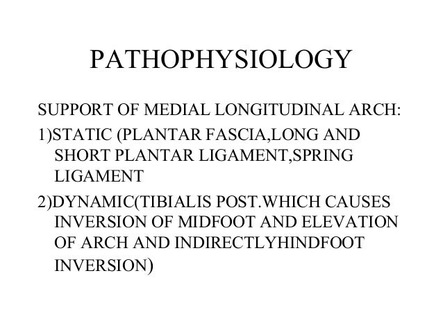
Causes
Flat feet causes greater pressure on the posterior tibial tendon than normal. As the person with flat feet ages, the muscles, tendons and ligaments weaken. Blood supplies diminish as arteries narrow. These conditions are magnified for obese patients because of their increased weight and atherosclerosis. Finally, the tendon gives out or tears. Most of the time, this is a slow process. Once the posterior tibial tendon and ligaments stretch, body weight causes the bones of the arch to move out of position. The foot rotates inward (pronation), the heel bone is tilted to the inside, and the arch appears collapsed. In some cases, the deformity progresses until the foot dislocates outward from the ankle joint.
Symptoms
Pain and swelling behind the inside of your ankle and along your instep. You may be tender behind the inner ankle where the posterior tibial tendon courses and occasionally get burning, shooting, tingling or stabbing pain as a result of inflammation of the nerve inside the tarsal tunnel. Difficulty walking, the inability to walk long distances and a generalised ache while walking even short distances. This may probably become more pronounced at the end of each day. Change in foot shape, sometimes your tendon stretches out, this is due to weakening of the tendon and ligaments. When this occurs, the arch in your foot flattens and a flatfoot deformity occurs, presenting a change in foot shape. Inability to tip-toe, a way of diagnosing Posterior Tibial Tendon Dysfunction is difficulty or inability to ?heel rise? (stand on your toes on one foot). Your tibialis posterior tendon enables you to perform this manoeuvre effectively. You may also experience pain upon attempting to perform a heel rise.
Diagnosis
Starting from the knee down, check for any bowing of the tibia. A tibial varum will cause increased medial stress on the foot and ankle. This is essential to consider in surgical planning. Check the gastrocnemius muscle and Achilles complex via a straight and bent knee check for equinus. If the range of motion improves to at least neutral with bent knee testing of the Achilles complex, one may consider a gastrocnemius recession. If the Achilles complex is still tight with bent knee testing, an Achilles lengthening may be necessary. Check the posterior tibial muscle along its entire course. Palpate the muscle and observe the tendon for strength with a plantarflexion and inversion stress test. Check the flexor muscles for strength in order to see if an adequate transfer tendon is available. Check the anterior tibial tendon for size and strength.
Non surgical Treatment
Treating PTTD is almost always easier the earlier you catch it. So, the first step in treatment is to see your doctor as soon as you begin experiencing painful symptoms. However, once your condition has been diagnosed, your podiatrist will likely try to give the upset tendon a bit of a break so it?ll calm down and stop being so painful. This can often be accomplished by immobilizing the foot using tape and padding, braces, or casts, depending on what your podiatrist believes will work best for you, and depending on the severity of your condition. You may also be instructed to reduce inflammation by applying ice to the area (usually 40 minutes on and 20 minutes off, with a thin towel between you and the ice). Or, you might take anti-inflammatory medications like ibuprofen (steroidal anti-inflammatory meds are actually likely to make this problem worse, and are not usually recommended in treating PTTD), or use ultrasound therapy. Once the inflammation has gone down a bit, your podiatrist may recommend using orthotics (prescription shoe inserts) to support your damaged arch. Ankle braces can also be helpful. 
Surgical Treatment
For those patients with PTTD that have severe deformity or have not improved with conservative treatments, surgery may be necessary to return them to daily activity. Surgery for PTTD may include repair of the diseased tendon and possible tendon transfer to a nearby healthy tendon, surgery on the surrounding bones or joints to prevent biomechanical abnormalities that may be a contributing factor or both.
Heel Pain And Discomfort Everything You Might Want To Understand Heel Serious Pain
Overview

Does your heel feel painful when you get up from bed every morning, or when you get up after sitting down for a long period of time? If you often experience this sort of pain, known as first step pain, you could be suffering from plantar fasciitis (pronounced fash-ee-eye-tus), an inflammation of the plantar fascia, or the band of muscle under the foot. Plantar fasciitis is the most common cause of Heel Pain.
Causes
Achilles tendon rupture, the tendon of the heel cord behind the ankle is torn. Bone bruise. Bone cyst, a solitary fluid-filled cyst (cavity) in a bone. Gout, levels of uric acid in the blood rise until the level becomes excessive (hyperuricemia), causing urate crystals to build up around the joints. This causes inflammation and severe pain when a gout attack happens. Neuroma (Morton's neuroma) a swollen nerve in the ball of the foot, commonly between the base of the second and third toes. Osteomyelitis , osteomyelitis means infection of the bone or bone marrow; inflammation of the bone due to infection. Osteomyelitis sometimes occurs as a complication of injury or surgery. In some cases, the infection may get into bone tissue from the bloodstream. Patients with osteomyelitis typically experience deep pain and muscle spasms in the inflammation area, as well as fever. Peripheral neuropathy, neuropathy is a collection of disorders that occurs when nerves of the peripheral nervous system (the part of the nervous system outside of the brain and spinal cord) are damaged. The condition is generally referred to as peripheral neuropathy, and it is most commonly due to damage to nerve axons. Neuropathy usually causes pain and numbness in the hands and feet. It can result from traumatic injuries, infections, metabolic disorders and exposure to toxins. One of the most common causes of neuropathy is diabetes. Problems with your gait, wrong posture when walking/running. Rheumatoid arthritis, rheumatoid arthritis, sometimes referred to as rheumatoid disease, is a chronic (long lasting), progressive and disabling auto-immune disease condition that causes inflammation and pain in the joints, the tissue around the joints, and other organs in the human body. Rheumatoid arthritis usually affects the joints in the hands and feet first, but any joint may become affected. Patients with rheumatoid arthritis commonly have stiff joints and feel generally unwell and tired.
Symptoms
Usually worse with the first few steps in the morning or at the initial point of activity. The latter usually gets better with continued activity (squeaky hinge analogy). Walking, running, sprinting, hill running and jumping will increase the pain. Often, the natural response is to walk on the outside of the foot - in supination - to lessen the stress on the plantar fascia - resulting in new problems.
Diagnosis
Your doctor will listen to your complaints about your heel and examine you to see what is causing the pain, and whether anything else has started it off. If the cause of your pain seems obvious, your doctor may be happy to start treatment straight away. However, some tests may be helpful in ruling out other problems. Blood tests may be done for arthritis. An Xray will show any arthritis in the ankle or subtalar joint, as well as any fracture or cyst in the calcaneum. (It will also show a spur if you have one, but as we know this is not the cause of the pain.) Occasionally a scan may be used to help spot arthritis or a stress fracture.
Non Surgical Treatment
Orthotics, by treating the cause of the problem, lead to the cure bette than 90% of the time. A small number of patients have waited so long that the plantar fascia has become thickened and filled with scar tissue and are not helped by ?conventional? means. Those are the patients that have, traditionally, required surgical treatment in which the plantar fascia is cut off the heel bone. Luckily, most surgery has been replaced by a relatively new mode of treatment, ESWT or Extracorporeal Shockwave Therapy. ESWT involves the application of multiple shockwaves to the diseased tendon or ligament and has an approximately 85 to 90% success rate. Keep in mind we are talking about this success rate in patients who are ?tough cases,? that is, already had the conventional treatment. The ESWT machines look like miniature renal lithtripsors (kidney stone crushers). There are virtually no side effects to ESWT other than the price as only about 30% of insurance companies are paying for it. They realize that it is less costly and safer than surgery but also know that many more people who would avoid surgery would have no problem getting ESWT so the volume of services would go up. You don?t have to live with painful heels.
Surgical Treatment
Although most patients with plantar fasciitis respond to non-surgical treatment, a small percentage of patients may require surgery. If, after several months of non-surgical treatment, you continue to have heel pain, surgery will be considered. Your foot and ankle surgeon will discuss the surgical options with you and determine which approach would be most beneficial for you. No matter what kind of treatment you undergo for plantar fasciitis, the underlying causes that led to this condition may remain. Therefore, you will need to continue with preventive measures. Wearing supportive shoes, stretching, and using custom orthotic devices are the mainstay of long-term treatment for plantar fasciitis.
Prevention

It is not always possible to prevent heel pain, but there are measures you can take to help avoid further episodes. Being overweight can place excess pressure and strain on your feet, particularly on your heels. This increases the risk of damaging your feet and heels. If you are overweight, losing weight and maintaining a healthy weight by combining regular exercise with a healthy, balanced diet can be beneficial for your feet. You can calculate your body mass index (BMI) to find out whether you are a healthy weight for your height and build. To work out your BMI, divide your weight in kilograms by your height in metres squared. A BMI of less than 18.5 means that you are underweight, 18.5-24.9 means that your weight is healthy, 25-29 means that you are overweight, 30-40 means that you are obese, over 40 means that you are morbidly obese. You can also use the BMI healthy weight calculator to work out your BMI.
Does your heel feel painful when you get up from bed every morning, or when you get up after sitting down for a long period of time? If you often experience this sort of pain, known as first step pain, you could be suffering from plantar fasciitis (pronounced fash-ee-eye-tus), an inflammation of the plantar fascia, or the band of muscle under the foot. Plantar fasciitis is the most common cause of Heel Pain.
Causes
Achilles tendon rupture, the tendon of the heel cord behind the ankle is torn. Bone bruise. Bone cyst, a solitary fluid-filled cyst (cavity) in a bone. Gout, levels of uric acid in the blood rise until the level becomes excessive (hyperuricemia), causing urate crystals to build up around the joints. This causes inflammation and severe pain when a gout attack happens. Neuroma (Morton's neuroma) a swollen nerve in the ball of the foot, commonly between the base of the second and third toes. Osteomyelitis , osteomyelitis means infection of the bone or bone marrow; inflammation of the bone due to infection. Osteomyelitis sometimes occurs as a complication of injury or surgery. In some cases, the infection may get into bone tissue from the bloodstream. Patients with osteomyelitis typically experience deep pain and muscle spasms in the inflammation area, as well as fever. Peripheral neuropathy, neuropathy is a collection of disorders that occurs when nerves of the peripheral nervous system (the part of the nervous system outside of the brain and spinal cord) are damaged. The condition is generally referred to as peripheral neuropathy, and it is most commonly due to damage to nerve axons. Neuropathy usually causes pain and numbness in the hands and feet. It can result from traumatic injuries, infections, metabolic disorders and exposure to toxins. One of the most common causes of neuropathy is diabetes. Problems with your gait, wrong posture when walking/running. Rheumatoid arthritis, rheumatoid arthritis, sometimes referred to as rheumatoid disease, is a chronic (long lasting), progressive and disabling auto-immune disease condition that causes inflammation and pain in the joints, the tissue around the joints, and other organs in the human body. Rheumatoid arthritis usually affects the joints in the hands and feet first, but any joint may become affected. Patients with rheumatoid arthritis commonly have stiff joints and feel generally unwell and tired.
Symptoms
Usually worse with the first few steps in the morning or at the initial point of activity. The latter usually gets better with continued activity (squeaky hinge analogy). Walking, running, sprinting, hill running and jumping will increase the pain. Often, the natural response is to walk on the outside of the foot - in supination - to lessen the stress on the plantar fascia - resulting in new problems.
Diagnosis
Your doctor will listen to your complaints about your heel and examine you to see what is causing the pain, and whether anything else has started it off. If the cause of your pain seems obvious, your doctor may be happy to start treatment straight away. However, some tests may be helpful in ruling out other problems. Blood tests may be done for arthritis. An Xray will show any arthritis in the ankle or subtalar joint, as well as any fracture or cyst in the calcaneum. (It will also show a spur if you have one, but as we know this is not the cause of the pain.) Occasionally a scan may be used to help spot arthritis or a stress fracture.
Non Surgical Treatment
Orthotics, by treating the cause of the problem, lead to the cure bette than 90% of the time. A small number of patients have waited so long that the plantar fascia has become thickened and filled with scar tissue and are not helped by ?conventional? means. Those are the patients that have, traditionally, required surgical treatment in which the plantar fascia is cut off the heel bone. Luckily, most surgery has been replaced by a relatively new mode of treatment, ESWT or Extracorporeal Shockwave Therapy. ESWT involves the application of multiple shockwaves to the diseased tendon or ligament and has an approximately 85 to 90% success rate. Keep in mind we are talking about this success rate in patients who are ?tough cases,? that is, already had the conventional treatment. The ESWT machines look like miniature renal lithtripsors (kidney stone crushers). There are virtually no side effects to ESWT other than the price as only about 30% of insurance companies are paying for it. They realize that it is less costly and safer than surgery but also know that many more people who would avoid surgery would have no problem getting ESWT so the volume of services would go up. You don?t have to live with painful heels.
Surgical Treatment
Although most patients with plantar fasciitis respond to non-surgical treatment, a small percentage of patients may require surgery. If, after several months of non-surgical treatment, you continue to have heel pain, surgery will be considered. Your foot and ankle surgeon will discuss the surgical options with you and determine which approach would be most beneficial for you. No matter what kind of treatment you undergo for plantar fasciitis, the underlying causes that led to this condition may remain. Therefore, you will need to continue with preventive measures. Wearing supportive shoes, stretching, and using custom orthotic devices are the mainstay of long-term treatment for plantar fasciitis.
Prevention
It is not always possible to prevent heel pain, but there are measures you can take to help avoid further episodes. Being overweight can place excess pressure and strain on your feet, particularly on your heels. This increases the risk of damaging your feet and heels. If you are overweight, losing weight and maintaining a healthy weight by combining regular exercise with a healthy, balanced diet can be beneficial for your feet. You can calculate your body mass index (BMI) to find out whether you are a healthy weight for your height and build. To work out your BMI, divide your weight in kilograms by your height in metres squared. A BMI of less than 18.5 means that you are underweight, 18.5-24.9 means that your weight is healthy, 25-29 means that you are overweight, 30-40 means that you are obese, over 40 means that you are morbidly obese. You can also use the BMI healthy weight calculator to work out your BMI.